Advanced Search
Volume 38 Issue 1
Jul.  2020
Turn off MathJax
Article Contents

TaiHeng SHI, Geng WU, Rong LI. Structures of Siliceous Hot Spring Deposits and Mechanism of Microbial Mineralization and Diagenesis[J]. Acta Sedimentologica Sinica, 2020, 38(1): 113-123. doi: 10.14027/j.issn.1000-0550.2019.023
Citation: TaiHeng SHI, Geng WU, Rong LI. Structures of Siliceous Hot Spring Deposits and Mechanism of Microbial Mineralization and Diagenesis[J]. Acta Sedimentologica Sinica, 2020, 38(1): 113-123. doi: 10.14027/j.issn.1000-0550.2019.023

Structures of Siliceous Hot Spring Deposits and Mechanism of Microbial Mineralization and Diagenesis

doi: 10.14027/j.issn.1000-0550.2019.023
Funds:

The Fundamental Research Funds for the Central Universities, China University of Geosciences (Wuhan) 2018319048

  • Received Date: 2018-12-28
  • Rev Recd Date: 2019-02-22
  • Publish Date: 2020-07-13
  • Hot springs are examples of continuous deposition in extreme environments, thus exploring the mineralization of hot spring microbes helps in understanding the Earth's ancient environment. Modern siliceous sinter is formed by both biological and non-biological interaction: the main mineral is Opal-A, with spicule, column and cluster etc. sedimentary structures. Petrographic studies of modern siliceous sinter are a good foundation for the study of the microbial mineralization process. In addition, it is significant for such studies to know more about the microbial mineralization process and the dissolution and re-deposition processes of opal diagenesis. At present, the study of microbial mineralization processes is flourishing. Specifically, microbially induced mineralization and microbially controlled mineralization are the two major mineralization processes, but there are many microbial mineralization mechanisms yet to be discovered. Therefore, examining mineralization by hot spring microbes is highly significant for strengthening our understanding of the formation and transformation of hot spring deposits.
  • [1] Vaughan R G, Hook S J, Calvin W M, et al. Surface mineral mapping at steamboat springs, Nevada, USA, with multiwavelength thermal infrared images[J]. Remote Sensing of Environment, 2005, 99(1/2):140-158. http://www.wanfangdata.com.cn/details/detail.do?_type=perio&id=8d551fdaf42fb932907adf7a69feea81
    [2] Pentecost A, Jones B, Renaut R W. What is a hot spring?[J]. Canadian Journal of Earth Sciences, 2003, 40(11):1443-1446. doi:  10.1139/e03-083
    [3] Smith I J, Lynne B Y, Jaworowski C, et al. The formation of geyser eggs at Old Faithful Geyser, Yellowstone National Park, U. S. A.[J]. Geothermics, 2018, 75:105-121. doi:  10.1016/j.geothermics.2018.04.006
    [4] 朱梅湘.地热系统的成岩成矿作用[J].矿物岩石地球化学通讯, 1993, 12(4):227-230. http://www.cnki.com.cn/Article/CJFDTotal-KYDH199304019.htm

    Zhu Meixiang. Diagenesis and mineralization of geothermal system[J]. Bulletin of Mineralogy, Petrology and Geochemistry, 1993, 12(4):227-230. http://www.cnki.com.cn/Article/CJFDTotal-KYDH199304019.htm
    [5] Guidry S A, Chafetz H S. Anatomy of siliceous hot springs:Examples from Yellowstone National Park, Wyoming, USA[J]. Sedimentary Geology, 2003, 157(1/2):71-106. doi:  10.1016-S0037-0738(02)00195-1/
    [6] Jones B, Renaut R W. Petrography and genesis of spicular and columnar geyserite from the Whakarewarewa and Orakeikorako geothermal areas, North Island, New Zealand[J]. Canadian Journal of Earth Sciences, 2003, 40(11):1585-1610. doi:  10.1139/e03-062
    [7] Walter M R. Chapter 3.3 Geyserites of Yellowstone National Park:An example of abiogenic "Stromatolites"[J]. Developments in Sedimentology, 1976, 20:87-112. https://www.sciencedirect.com/science/article/pii/S0070457108711312
    [8] Jones B, Renaut R W, Rosen M R. Microbial construction of siliceous stalactites at geysers and hot springs:Examples from the Whakarewarewa geothermal area, North Island, New Zealand[J]. Palaios, 2001, 16(1):73-94. doi:  10.1669/0883-1351(2001)016<0073:MCOSSA>2.0.CO;2
    [9] Channing A, Edwards D, Sturtevant S. A geothermally influenced wetland containing unconsolidated geochemical sediments[J]. Canadian Journal of Earth Sciences, 2004, 41(7):809-827. doi:  10.1139/e04-034
    [10] Djokic T, van Kranendonk M J, Campbell K A, et al. Earliest signs of life on land preserved in ca. 3.5 Ga hot spring deposits[J]. Nature Communications, 2017, 8:15263. doi:  10.1038/ncomms15263
    [11] Jones B, Renaut R W, Owen R B. Life cycle of a geyser discharge apron:Evidence from Waikite geyser, Whakarewarewa geothermal area, North Island, New Zealand[J]. Sedimentary Geology, 2011, 236(1/2):77-94.
    [12] 李红中, 周永章, 杨志军, 等.秦岭造山带西段八方山-二里河地区硅质岩的地球化学特征及其地质意义[J].岩石学报, 2009, 25(11):3094-3102. http://d.old.wanfangdata.com.cn/Periodical/ysxb98200911035

    Li Hongzhong, Zhou Yongzhang, Yang Zhijun, et al. Geochemical characteristics and their geological implications of cherts from Bafangshan-Erlihe area in western Qinling orogen[J]. Acta Petrologica Sinica, 2009, 25(11):3094-3102. http://d.old.wanfangdata.com.cn/Periodical/ysxb98200911035
    [13] 何俊国.特提斯构造域东段中、新生代硅质岩及其沉积环境研究[D].广州: 中山大学, 2009.

    He Junguo. Study on Mesozonic and Caenozonic chert and its sedimentary environment in Tethyan Domain[D]. Guangzhou: Sun Yat-sen University, 2009.
    [14] Fortney N W, He S, Converse B J, et al. Microbial Fe (III) oxide reduction potential in chocolate pots hot spring, Yellowstone National Park[J]. Geobiology, 2016, 14(3):255-275. doi:  10.1111/gbi.12173
    [15] Day R, Jones B. Variations in water content in opal-A and opal-CT from geyser discharge aprons[J]. Journal of Sedimentary Research, 2008, 78(4):301-315. doi:  10.2110/jsr.2008.030
    [16] Jones B, Renaut R W. Water content of opal-A:Implications for the origin of laminae in geyserite and sinter[J]. Journal of Sedimentary Research, 2004, 74(1):117-128. doi:  10.1306/052403740117
    [17] Dunckel A E, Cardenas M B, Sawyer A H, et al. High-resolution in-situ thermal imaging of microbial mats at El Tatio Geyser, Chile shows coupling between community color and temperature[J]. Geophysical Research Letters, 2009, 36(23):L23403. doi:  10.1029/2009GL041366
    [18] Boudreau A E, Lynne B Y. The growth of siliceous sinter deposits around high-temperature eruptive hot springs[J]. Journal of Volcanology and Geothermal Research, 2012, 247-248:1-8. doi:  10.1016/j.jvolgeores.2012.07.008
    [19] Garcia-Valles M, Fernandez-Turiel J L, Gimeno-Torrente D, et al. Mineralogical characterization of silica sinters from the El Tatio geothermal field, Chile[J]. American Mineralogist, 2008, 93(8/9):1373-1383. http://www.wanfangdata.com.cn/details/detail.do?_type=perio&id=10.2138/am.2008.2583
    [20] Rodgers K A, Browne P R L, Buddle T F, et al. Silica phases in sinters and residues from geothermal fields of New Zealand[J]. Earth Science Reviews, 2004, 66(1/2):1-61. http://cn.bing.com/academic/profile?id=d6c616023cb274788ed2a9f61dbd4060&encoded=0&v=paper_preview&mkt=zh-cn
    [21] Jones B, Renaut R W. Hot spring and geyser sinters:The integrated product of precipitation, replacement, and deposition[J]. Canadian Journal of Earth Sciences, 2003, 40(11):1549-1569. doi:  10.1139/e03-078
    [22] Smith D J, Jenkin G R T, Petterson M G, et al. Unusual mixed silica-carbonate deposits from magmatic-hydrothermal hot springs, Savo, Solomon Islands[J]. Journal of the Geological Society, 2011, 168:1297-1310. doi:  10.1144/0016-76492011-003
    [23] Kim J W, Kogure T, Yang K, et al. The characterization of CaCO 3 in a geothermal environment:A SEM/TEM-EELS study[J]. Clays and Clay Minerals, 2012, 60(5):484-495. doi:  10.1346/CCMN.2012.0600505
    [24] Watts-Henwood N, Campbell K A, Lynne B Y, et al. Snapshot of hot-spring sinter at Geyser Valley, Wairakei, New Zealand, following anthropogenic drawdown of the geothermal reservoir[J]. Geothermics, 2017, 68:94-114. doi:  10.1016/j.geothermics.2017.03.002
    [25] Braunstein D, Lowe D R. Relationship between spring and geyser activity and the deposition and morphology of high temperature (> 73℃) siliceous sinter, Yellowstone National Park, Wyoming, U. S. A.[J]. Journal of Sedimentary Research, 2001, 71(5):747-763. doi:  10.1306/2DC40965-0E47-11D7-8643000102C1865D
    [26] Jones B, Renaut R W. Formation of silica oncoids around geysers and hot springs at El Tatio, northern Chile[J]. Sedimentology, 1997, 44(2):287-304. doi:  10.1111/j.1365-3091.1997.tb01525.x
    [27] Renaut R W, Jones B, Tiercelin J J, et al. Sublacustrine precipitation of hydrothermal silica in rift lakes:Evidence from Lake Baringo, central Kenya Rift Valley[J]. Sedimentary Geology, 2002, 148(1/2):235-257. doi:  10.1016-S0037-0738(01)00220-2/
    [28] Jones B, Renaut R W, Rosen M R. Microbial biofacies in hot-spring sinters:A model based on Ohaaki Pool, North Island, New Zealand[J]. Journal of Sedimentary Research, 1998, 68(3):413-434. doi:  10.2110/jsr.68.413
    [29] Jones B, Renaut R W, Rosen M R. Taphonomy of silicified filamentous microbes in modern geothermal sinters:Implications for identification[J]. Palaios, 2001, 16(6):580-592. doi:  10.1669/0883-1351(2001)016<0580:TOSFMI>2.0.CO;2
    [30] Handley K M, Turner S J, Campbell K A, et al. Silicifying biofilm exopolymers on a hot-spring microstromatolite:Templating nanometer-thick laminae[J]. Astrobiology, 2008, 8(4):747-770. doi:  10.1089/ast.2007.0172
    [31] Rimstidt J D, Cole D R. Geothermal mineralization; I, the mechanism of formation of the Beowawe, Nevada, siliceous sinter deposit[J]. American Journal of Science, 1983, 283(8):861-875. doi:  10.2475/ajs.283.8.861
    [32] Guidry S A, Chafetz H S. Factors governing subaqueous siliceous sinter precipitation in hot springs:Examples from Yellowstone National Park, USA[J]. Sedimentology, 2002, 49(6):1253-1267. doi:  10.1046/j.1365-3091.2002.00494.x
    [33] Renaut R W, Owen R B, Ego J K. Geothermal activity and hydrothermal mineral deposits at southern Lake Bogoria, Kenya rift valley:Impact of lake level changes[J]. Journal of African Earth Sciences, 2017, 129:623-646. doi:  10.1016/j.jafrearsci.2017.01.012
    [34] Ruff S W, Farmer J D. Silica deposits on Mars with features resembling hot spring biosignatures at El Tatio in Chile[J]. Nature Communications, 2016, 7:13554. doi:  10.1038/ncomms13554
    [35] Lewis A J, Palmer M R, Sturchio N C, et al. The rare earth element geochemistry of acid-sulphate and acid-sulphatechloride geothermal systems from Yellowstone National Park, Wyoming, USA[J]. Geochimica et Cosmochimica Acta, 1997, 61(4):695-706. doi:  10.1016/S0016-7037(96)00384-5
    [36] Jones B, Renaut R W. Influence of thermophilic bacteria on calcite and silica precipitation in hot springs with water temperatures above 90℃:Evidence from Kenya and New Zealand[J]. Canadian Journal of Earth Sciences, 1996, 33(1):72-83. doi:  10.1139/e96-008
    [37] Jones B, Renaut R W, Rosen M R. Silicified microbes in a geyser mound:The enigma of low-temperature cyanobacteria in a high-temperature setting[J]. Palaios, 2003, 18(2):87-109. doi:  10.1669/0883-1351(2003)18<87:SMIAGM>2.0.CO;2
    [38] Lowe D R, Braunstein D. Microstructure of high-temperature (>73℃) siliceous sinter deposited around hot springs and geysers, Yellowstone National Park:The role of biological and abiological processes in sedimentation[J]. Canadian Journal of Earth Sciences, 2003, 40(11):1611-1642. doi:  10.1139/e03-066
    [39] Jones B, Renaut R W, Rosen M R, et al. Coniform stromatolites from geothermal systems, North Island, New Zealand[J]. Palaios, 2002, 17(1):84-103. doi:  10.1669/0883-1351(2002)017<0084:CSFGSN>2.0.CO;2
    [40] Guidry S A, Chafetz H S. Depositional facies and diagenetic alteration in a relict siliceous hot-spring accumulation:Examples from Yellowstone National Park, U. S. A.[J]. Journal of Sedimentary Research, 2003, 73(5):806-823. doi:  10.1306/022803730806
    [41] Campbell K A, Guido D M, Gautret P, et al. Geyserite in hot-spring siliceous sinter:Window on earth's hottest terrestrial (Paleo) environment and its extreme life[J]. Earth-Science Reviews, 2015, 148:44-64. doi:  10.1016/j.earscirev.2015.05.009
    [42] Jones B, Renaut R W, Rosen M R. Stromatolites forming in acidic hot-spring waters, North Island, New Zealand[J]. Palaios, 2000, 15(5):450-475. doi:  10.1669/0883-1351(2000)015<0450:SFIAHS>2.0.CO;2
    [43] Barbieri R, Cavalazzi B, Stivaletta N, et al. Silicified biota in high-altitude, geothermally influenced ignimbrites at El Tatio geyser field, Andean Cordillera (Chile)[J]. Geomicrobiology Journal, 2014, 31(6):493-508. doi:  10.1080/01490451.2013.836691
    [44] 付伟, 霍晓萍, 周永章, 等.太古宙的生命起源信息及研究评述[J].中山大学研究生学刊(自然科学、医学版), 2006, 27(2):42-49. http://kns.cnki.net/KCMS/detail/detail.aspx?dbcode=CJFD&filename=YJSK200602006

    Fu Wei, Huo Xiaoping, Zhou Yongzhang, et al. Progresses and reviews on the Archean life[J]. Journal of the Graduates Sun Yat-Sen University (Natural Sciences、Medicine), 2006, 27(2):42-49. http://kns.cnki.net/KCMS/detail/detail.aspx?dbcode=CJFD&filename=YJSK200602006
    [45] Labrenz M, Druschel G K, Thomsen-Ebert T, et al. Formation of sphalerite (ZnS) deposits in natural biofilms of sulfate-reducing bacteria[J]. Science, 2000, 290(5497):1744-1747. doi:  10.1126/science.290.5497.1744
    [46] 王海雷.西藏地热区微生物对铯的吸附及其对铯硅华成矿的贡献[D].北京: 中国地质科学院, 2006.

    Wang Hailei. Accumulation of cesium by microorganisms and role of microorganisms in the formation of cesium-bearing geyserite in geothermal areas, Tibet[D]. Beijing: Chinese Academy of Geological Sciences, 2006.
    [47] Joshi K B. Microbes:Mini iron factories[J]. Indian Journal of Microbiology, 2014, 54(4):483-485. doi:  10.1007/s12088-014-0497-1
    [48] 王海雷, 郑绵平, 黄晓星.菌株Thermus sp. TibetanG7对铯的吸附:热泉铯硅华形成过程中生物成矿作用的征兆[J].科学通报, 2007, 52(17):2043-2048. doi:  10.3321/j.issn:0023-074x.2007.17.012

    Wang Hailei, Zheng Mianping, Huang Xiaoxing. Cesium accumulation by bacterium Thermus sp. TibetanG7:Hints for biomineralization of cesium-bearing geyserite in hot springs in Tibet, China[J]. Chinese Science Bulletin, 2007, 52(17):2043-2048. doi:  10.3321/j.issn:0023-074x.2007.17.012
    [49] Peng X T, Zhou H Y, Li J T, et al. Intracellular and extracellular mineralization of a microbial community in the Edmond deep-sea vent field environment[J]. Sedimentary Geology, 2010, 229(4):193-206. doi:  10.1016/j.sedgeo.2010.06.003
    [50] 张天乐, 王宗良, 胡云中.腾冲现代热泉系统硅华的矿物学特征及其地质意义[J].岩石矿物学杂志, 1997, 16(2):170-178. http://www.cnki.com.cn/Article/CJFDTotal-YSKW702.007.htm

    Zhang Tianle, Wang Zongliang, Hu Yunzhong. Mineralogy of geyserite from the Tengchong active hot spring system and its geological implications[J]. Acta Petrologica et mineralogica, 1997, 16(2):170-178. http://www.cnki.com.cn/Article/CJFDTotal-YSKW702.007.htm
    [51] 张余.藏南中生代以来硅质沉积物的微组构特征及其地质意义[D].广州: 中山大学, 2007. http://www.wanfangdata.com.cn/details/detail.do?_type=degree&id=Y1086467

    Zhang Yu. Mirco-fabrics and their geological implications of silica sediments from southern Tibet (China)[D]. Guangzhou: Sun Yat-Sen University, 2007. http://www.wanfangdata.com.cn/details/detail.do?_type=degree&id=Y1086467
    [52] 赵元艺, 韩景仪, 郭立鹤, 等.西藏搭格架热泉型铯矿床矿物学与矿石组构特征及地质意义[J].岩石学报, 2008, 24(3):519-530. http://d.old.wanfangdata.com.cn/Periodical/ysxb98200803012

    Zhao Yuanyi, Han Jingyi, Guo Lihe, et al. Characteristics and geological significance of mineralogy and fabrics for the hot spring cesium deposit occurring within the Targejia district, Tibet[J]. Acta Petrologica Sinica, 2008, 24(3):519-530. http://d.old.wanfangdata.com.cn/Periodical/ysxb98200803012
    [53] 周永章, 付伟, 杨志军, 等.雅鲁藏布江缝合带及藏南地区硅质岩微组构特征及其地质意义[J].岩石学报, 2006, 22(3):742-750. http://d.old.wanfangdata.com.cn/Periodical/ysxb98200603024

    Zhou Yongzhang, Fu Wei, Yang Zhijun, et al. Microfabrics of chert from Yarlung Zangbo suture zone and southern Tibet and its geological implications[J]. Acta Petrologica Sinica, 2006, 22(3):742-750. http://d.old.wanfangdata.com.cn/Periodical/ysxb98200603024
    [54] Lynne B Y, Campbell K A. Morphologic and mineralogic transitions from opal-A to opal-CT in low-temperature siliceous sinter diagenesis, Taupo Volcanic Zone, New Zealand[J]. Journal of Sedimentary Research, 2004, 74(4):561-579. doi:  10.1306/011704740561
    [55] Jones B, Renaut R W. Microstructural changes accompanying the opal-A to opal-CT transition:New evidence from the siliceous sinters of Geysir, Haukadalur, Iceland[J]. Sedimentology, 2007, 54(4):921-948. doi:  10.1111/j.1365-3091.2007.00866.x
    [56] 周永章, 付伟, 杨志军, 等.藏南硅质岩及富SiO2热水流体成岩成矿作用研究[J].矿物岩石地球化学通报, 2008, 27(增刊1):351-352. http://d.old.wanfangdata.com.cn/Periodical/kwysdqhxtb2008z1190

    Zhou Yongzhang, Fu Wei, Yang Zhijun, et al. Study on diagenesis and mineralization of cherts and Si-rich hydrothermal fluids in southern Tibet[J]. Bulletin of Mineralogy, Petrology and Geochemistry, 2008, 27(Suppl. 1):351-352. http://d.old.wanfangdata.com.cn/Periodical/kwysdqhxtb2008z1190
    [57] Watanabe S, Hayashi K I. Mineralogy, sulfur isotope and fluid inclusion studies of hydrothermal ore at the Hakurei Deposit, Bayonnaise Knoll, Izu-Bonin Arc[J]. Resource Geology, 2014, 64(2):77-90. doi:  10.1111/rge.12029
    [58] Konhauser K O. Diversity of bacterial iron mineralization[J]. Earth-Science Reviews, 1998, 43(3/4):91-121. doi:  10.1016-S0012-8252(97)00036-6/
    [59] Tivey M K, Stakes D S, Cook T L, et al. A model for growth of steep-sided vent structures on the Endeavour Segment of the Juan de Fuca Ridge:Results of a petrologic and geochemical study[J]. Journal of Geophysical Research:Solid Earth, 1999, 104(B10):22859-22883.
    [60] Georgieva M N, Little C T S, Ball A D, et al. Mineralization of Alvinella polychaete tubes at hydrothermal vents[J]. Geobiology, 2015, 13(2):152-169. doi:  10.1111/gbi.12123
    [61] Tivey M K, Delaney J R. Growth of large sulfide structures on the endeavour segment of the Juan de Fuca ridge[J]. Earth and Planetary Science Letters, 1986, 77(3/4):303-317. http://cn.bing.com/academic/profile?id=b72570dc7c3b2d32bbb336777ecb8d2e&encoded=0&v=paper_preview&mkt=zh-cn
    [62] Jones B, de Ronde C E J, Renaut R W. Mineralized microbes from Giggenbach submarine volcano[J]. Journal of Geophysical Research:Solid Earth, 2008, 113(B8):B08S05. http://www.wanfangdata.com.cn/details/detail.do?_type=perio&id=10.1029/2007JB005482
    [63] Rasmussen B, Muhling J R, Suvorova A, et al. Greenalite precipitation linked to the deposition of banded iron formations downslope from a late Archean carbonate platform[J]. Precambrian Research, 2017, 290:49-62. doi:  10.1016/j.precamres.2016.12.005
  • 加载中
通讯作者: 陈斌, bchen63@163.com
  • 1. 

    沈阳化工大学材料科学与工程学院 沈阳 110142

  1. 本站搜索
  2. 百度学术搜索
  3. 万方数据库搜索
  4. CNKI搜索

Figures(5)

Article Metrics

Article views(627) PDF downloads(178) Cited by()

Proportional views
Related
Publishing history
  • Received:  2018-12-28
  • Revised:  2019-02-22
  • Published:  2020-07-13

Structures of Siliceous Hot Spring Deposits and Mechanism of Microbial Mineralization and Diagenesis

doi: 10.14027/j.issn.1000-0550.2019.023
Funds:

The Fundamental Research Funds for the Central Universities, China University of Geosciences (Wuhan) 2018319048

Abstract: Hot springs are examples of continuous deposition in extreme environments, thus exploring the mineralization of hot spring microbes helps in understanding the Earth's ancient environment. Modern siliceous sinter is formed by both biological and non-biological interaction: the main mineral is Opal-A, with spicule, column and cluster etc. sedimentary structures. Petrographic studies of modern siliceous sinter are a good foundation for the study of the microbial mineralization process. In addition, it is significant for such studies to know more about the microbial mineralization process and the dissolution and re-deposition processes of opal diagenesis. At present, the study of microbial mineralization processes is flourishing. Specifically, microbially induced mineralization and microbially controlled mineralization are the two major mineralization processes, but there are many microbial mineralization mechanisms yet to be discovered. Therefore, examining mineralization by hot spring microbes is highly significant for strengthening our understanding of the formation and transformation of hot spring deposits.

TaiHeng SHI, Geng WU, Rong LI. Structures of Siliceous Hot Spring Deposits and Mechanism of Microbial Mineralization and Diagenesis[J]. Acta Sedimentologica Sinica, 2020, 38(1): 113-123. doi: 10.14027/j.issn.1000-0550.2019.023
Citation: TaiHeng SHI, Geng WU, Rong LI. Structures of Siliceous Hot Spring Deposits and Mechanism of Microbial Mineralization and Diagenesis[J]. Acta Sedimentologica Sinica, 2020, 38(1): 113-123. doi: 10.14027/j.issn.1000-0550.2019.023
  • 热泉是大气水循环到浅层地壳深部[1]后,经岩浆驱动,从地表流出时水温≥36.7 ℃的泉[2]。热泉中矿物沉积形成热泉沉积物[3]。依据矿物组成,可将热泉沉积物划分为硅华(主要矿物为Opal-A,Opal-CT, 石英等)、钙华(主要矿物为方解石,文石)、盐华(盐类矿物)和硫华[4]

    目前热泉沉积研究主要分为:岩石学研究,生物学研究,地球化学研究及微生物矿化研究四个领域,其中以岩石学研究为基础,生物学及地球化学研究相继发展,微生物矿化研究最热门。

    热泉沉积岩石学研究始于热泉研究之初,早先该领域的研究内容主要在于解译与古代岩石中相似沉积特征[5],寻找热泉沉积中残存的微生物痕迹,探索热泉沉积的成因[6, 7-9],逐渐达成热泉沉积是由生物与非生物因素共同作用形成这一共识。目前,单纯的岩相学研究已进入瓶颈期,现代热泉沉积岩石学研究趋向于寻找古老热泉沉积中残存的生物痕迹。例如,澳大利亚古老热泉叠层石中生物证据的发现将最早的陆地生命提前到3.5 Ga,使寻找古老的火星生命成为可能[10];热泉沉积可以记录热泉的喷发休眠史[11]等。

    热泉沉积的生物学及地球化学研究虽起步晚于岩石学研究,但发展十分迅速。早期热泉微生物方面的研究主要在于了解热泉中的微生物类群及分布,拓展对极端环境条件下微生物进化及生命特征的认识;地球化学研究早期则注重于研究古老及现代热泉沉积中的元素组成,来源及富集特点,进而反演古构造演化和推测硅质岩或硅华成因等[12-13]。然而,现代生物学研究多与地球化学研究相结合,探索元素在微生物地球化学循环中的转变及沉积特点,进而形成热泉微生物矿化作用研究领域。例如,Fe可参与微生物的地球化学循环而保存在热泉沉积中[14]等,促进了热泉微生物矿化领域的研究。

    本文以硅质热泉沉积为例,针对热泉微生物矿化作用涉及岩石学、生物学和地球化学等过程的特点,系统总结了硅华沉积体系的组成、硅华的沉积结构及其时空变化、微生物矿化及硅华的成岩转化过程,然后分析了现代热泉微生物矿化作用研究与热泉沉积成岩转化过程中存在的问题,结合最新的微生物矿化作用研究成果,对热泉微生物矿化作用的研究进行了展望。

  • 在热泉沉积研究中,温度、pH和宏观特征一般都会在野外测量完成,实验室工作主要在于确定热泉沉积物的岩石、矿物、元素及同位素特点,常用的方法主要包括:岩石薄片、扫描电镜、数字模型、X射线衍射、电子探针、ICP-MS和ICP-OES等,其中不乏改进和创新性的方法。

    在Opal水含量的研究中,Day et al. [15]巧妙地使用了电子探针及背散射电子成像技术,使用电子探针对Ca,Mg,Al,Na,Fe,Mn,K等能够结合O的元素含量进行测试,对这些元素含量低于百分之一的样品做Si和O的含量分析,两者按照1:2确定多余的O含量,进而计算出总水含量。该方法通常结合傅里叶变换红外光谱法使用,确定总水含量与不同类型的水含量,减小误差。背散射电子像只能反映同一个相片内的平均原子质量差异,不同相片间无法比较[16]。傅里叶变换红外光谱法虽能分析总水量和不同类型的水(H2O和OH-)含量,但测量精度不足[16]

    近年来,不同学者根据自己的需求在对热泉沉积进行研究时创新性地使用了多种方法,如Vaughan et al.[1]利用机载多光谱和高光谱热红外(TIR)图像数据,依据不同矿物对可见光及红外波的吸收特征,识别活动地热系统的地表矿物类型并绘制出地表矿物分布图,分析地表矿物的分布特征,但这种识别方式的空间分辨率相对较低。Dunckel et al. [17]使用热成像技术,将热泉温度变化与热泉微生物识别联系在一起,更加直观的表示出热泉微生物类群与温度的关系。Boudreau et al.[18]研究热泉沉积形成过程中蒸发与二氧化硅析出相互作用时,使用数字模型,设定一个初始的可光滑或粗糙的表面,输入热液硅浓度,蒸发速率等不同类型的参数后,通过程序算法得到与实际相符的沉积情况。Guidry et al.[5]将无菌的玻璃片放置于研究区,先进行原地模拟沉积,再将沉积物与早先形成的沉积做矿物组成及沉积结构的对比研究。

  • 硅华主要由胶状Opal-A组成[9, 19-20],多沉积于碱性热泉中[21],少见于酸性硫酸盐热泉[22]和硫酸盐氯化物热泉中[3]。次要矿物包括方解石、蒸发矿物(石膏等)、黏土矿物(高岭石,蒙脱石等)、石质碎屑、铁锰氧化物等,多沉积于硅华的孔隙或裂隙间[21, 23]

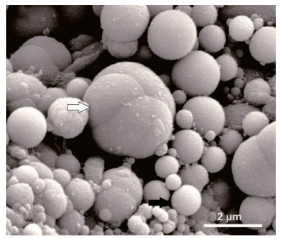
  • Opal-A,球形或椭球形(颗粒状)或聚成葡萄状(图 1),平均直径约5~10 µm,表面光滑。其沉积方式包括直接沉淀、以微生物作为模板进行交代和结壳沉积或作为胶结物沉积[21],通常沉积为层状。

    Figure 1.  SEM photomicrograph of particulate Opal-A(black arrow) and botryoidal clusters of Opal-A (white arrow) (modified from Smith et al.[3])

    Opal-A中水存在的形式包括H2O和OH- [21, 23-24],通常吸附于暴露的Opal表面或微孔中[23]。平均水含量高的Opal层是快速沉积的结果,反之则是慢速沉积的结果[23]

    Opal-A沉积的主要控制因素包括热液快速冷却[3, 18, 23, 25-26]、热液蒸发[7, 11, 18, 23]、pH改变[7, 27]、Opal-A与各种有机物及Fe和Mn等作为沉积核心和模板[23-24, 26, 28-30]、流体动力作用(如波浪等)[3, 25]、阳离子效应[3, 31]、脱气作用(CO2,CO)[32]和时间等。此外,生物因素对Opal-A的沉积也起到重要作用[32]

  • 独自沉积,或与Opal-A共生沉积[28],呈微晶、纤维状或鲕粒状[21],多沉积于靠近泉口处,离泉口较远处的水道和泉边缘的叠层石结构中也会有方解石沉积[21, 28]。沉积的主要原因在于快速的脱气作用(CO2,CO)[33]和过饱和沉积[34]。文石沉积的报道仅有一处[21]

  • 包括高岭石[9],蒙脱石,海泡石和皂石[1, 35]等。多包覆方解石或覆盖下伏层状Opal-A,部分填充及包覆微生物,通常呈纤维状或网状结构(推测为微生物形成,或由生物膜或细胞外聚合物捕获硅或其他元素后经脱水形成[36])。以富含Si,Mg,Al,K或Ca为特征[37],并且一般富铝的黏土矿物多集中在泉口。因为易受早期成岩作用影响,黏土矿物通常不与Opal-A呈夹层状沉积[21]

  • 包括石膏和盐岩,多发现于泉华和微叠层石中[5],自形的石膏晶体沉积在叠层石表面,嵌入Opal-A层或包覆微生物[1, 19]。自形盐岩多沉积在叠层石表面或泉华内裂缝,主要通过蒸发[19]或硫化物氧化沉积[21]

  • 石质碎屑分为内源碎屑和外源碎屑,主要是石英,长石和一些重矿物,主要沉积在孔隙中[3, 19, 38]。外源碎屑(水沉积,物质流,风沉积)来自流域内和谷坡的松散沉积颗粒,其丰富度和组成依赖于地势,土壤,母岩组分和气候(控制植物覆盖,径流,侵蚀速率),部分还包括火山碎屑[21, 39]。大多数内源碎屑来源于被侵蚀和改造的多孔易碎泉华,由喷孔形成或爆发式的喷发形成[21]。另外,地下水沉降、地震活动、风化过程、干湿交替作用、干燥的硅化微生物席和冻融作用[40]也会形成内源碎屑,多为棱角次棱角状。通常石质碎屑依重力分异沿泉口到泉边沉积,沉积在叠层石和针状等结构空隙间。

    微生物可以在生存或死亡后通过排水系统运移后沉积为生物碎屑[3]。生物碎屑还包括细枝、树叶、硅化和未硅化的生物残体等[19, 21, 36]

  • 硫,呈枝状晶体排列,填充缝隙,偶有发现在叠层石表面或嵌入Opal-A层中,多被氧化[21]。赤铁矿和锰的氧化物等发现于硅化的微生物细胞中[32],多沉积于好氧的环境条件下[40]。浅水的高温泉口环境下多沉积沸石[40]

    综上可以看出,硅质热泉沉积的主要矿物为Opal-A,其间夹杂着其他自生或搬运来的碎屑矿物沉积,沉积形态与沉积方式由热泉沉积环境或微生物控制。目前,热泉沉积物的矿物组成及来源的研究已十分清晰,通常作为热泉沉积其他领域研究的基础。

  • 硅质热泉沉积体系有两种分类方案。Guidry et al. [5]根据流体的温度将硅质热泉沉积体系划分为7个沉积亚相和间歇泉相:1)泉口亚相(Vent facies,> 95 ℃),2)近泉口亚相(Proximal vent facies,< 95 ℃),3)泉池亚相(Pool facies,约80 ℃~90 ℃),4)泉池边缘亚相(Poolmargin facies,约80 ℃),5)泉池旋涡亚相(Pool eddy facies,< 80 ℃),6)流径亚相(Discharge channel/flowpath facies,< 80 ℃),7)冲积平原亚相(Debris apron facies),8)间歇泉相(Geyser facies)。Braunstein et al.[25]和Lowe et al.[38]根据水动力条件(流体是否具有波浪、波浪幅度、是否沸腾),将硅质热泉沉积体系划分为6个沉积亚相:1)无波浪不沸腾泉亚相(Non-surging, non-boiling springs),2)微波浪不沸腾泉亚相(Gently surging, non-boiling springs),3)无波浪到微波浪沸腾泉亚相(Non-surging to gently surging, boiling springs),4)强波浪泉亚相(Strongly surging springs),5)间歇喷泉亚相(Fountain geysers),6)间歇锥形喷泉亚相(Cone geysers)。虽然依据水动力条件划分的沉积相更便于对沉积结构的形成进行研究,但依据流体温度划分的沉积相更易于野外的区分和辨别,而且有利于热泉微生物的区分,因此本文采用第一种划分方式介绍硅质热泉沉积体系的沉积亚相特征(图 2)。

    Figure 2.  Cross-section sketch of the silica hot spring sedimentary system (modified from Guidry et al. [5])

  • 多为非生物沉积[7]且主要为负延性玉髓(直径约0.5~0.75 cm)填充(图 2),晶体聚集成一系列凸起,纵截面呈天然的贝壳形,横截面呈菱形。水下玉髓晶体表面有一些颗粒状Opal-A[24-25]和葡萄状Opal-A沉积[5, 41]。部分高温泉口也会有较少嗜热菌产生的沉积[37]

  • 主要为颗粒状Opal-A沉积,内部通常中空,呈海绵状,多与正延性玉髓互层(图 2)或以Opal-A结壳的形式沉积。Opal层形态受下伏Opal-A层或近泉口底部微形态控制[41]。通常近泉口处沉积层最厚,部分近泉口处还沉积有硅质角砾岩[7]

  • 泉池深约5~10 cm,底部光滑,以涟漪状Opal为特征[5, 25],底部细沟的长轴指向流径方向。水下环境为颗粒状Opal-A沉积[1, 25, 31]或葡萄状沉积[3],泉池底部覆盖弱胶结的核形石,鲜有树枝状结构生长的锰的氧化物[6, 38, 40]和自生硫沉积[42]

    硅质核形石由Opal-A组成,外壳富微生物[3],含有少量砂级角砾状石英[5],内部多为同心层状[26, 28],少数夹有针状结构或簇丛状结构。Jones et al. [26]推测泉池底部的豆状结构或为核形石的前身。

  • 边缘相的沉积速率最快,以叠层石状的沉积结构最为常见,通常被认为是大量不同群落微生物的产物。叠层石结构约20~30 cm宽,2~3 cm高,表面为白色或因自生硫或明矾石沉积而显示灰色略带微黄,边缘和底部为白色粉末状Opal-A,内部由不规则的浅色层和富含有机质的深色层相间组成[25]。叠层石结构的孔隙(直径约1 mm)丰富,表面和孔隙多被微生物和黏液占据[5],部分叠层石结构的表面长有更细小的针状结构。冷却和蒸发作用是叠层石状结构形成的最主要的非生物因素,微生物或诱发Opal-A沉积或作为Opal-A沉积的模板[5]

    泉池的边缘因水动力条件变化大,除叠层石结构外,还沉积有较多的针状、柱状和簇丛状结构。针状结构通常1~3 cm高,直径0.5~1 mm,表面光滑或具有瘤状或树枝状修饰,横截面呈椭圆形,内部具有层状核心,外部包覆硅质结壳,间隙内发育有硅化的微生物席和树枝状结构[6]。相邻的针状结构或由间隙相隔或合并聚集成柱状结构或簇丛状结构[38]。针状、柱状和簇丛状结构的间隙发育有树枝状结构,该结构外层通常包覆Opal-A结壳,内部具有中空的孔隙,目前并不确定是否为微生物导致矿物呈枝状生长,但前人研究表明快速冷却和脱气作用能使硫、锰的氧化物[40]和方解石[6]等矿物呈树枝状生长。这三种结构多生长于泉水飞溅区和水池边缘、浅的流径边缘、间歇性暴露的盆缘和泄水道等不受飞溅水影响但水面震荡,蒸发冷却快的干湿交替区域[18, 25, 38]

    泉池陡峭边缘滴水处可沉积硅质石笋。硅质石笋彼此分离或相互连接,表面覆盖橙色、绿色、棕色微生物席,边缘多呈鳞片状[8],内部具有中空管,呈同心层状生长,层内具有多变的微孔[8]。在一些规模很小的小泉口的水池边缘,会形成平行于水气界面的指状结构,外形与手指相似,内部为层状结构[38]

  • 该沉积亚相通常难以辨别,具有约2~3 cm深的稳定水池(图 2),但因温度、pH和水平面的变化较大,Opal-A呈异化粒沉积,内部层状结构发育差,结合松散。硅化的化石结构丰富但少有内部细胞结构保留。该沉积亚相中还包括少量可能是风搬运的石英沉积,以Opal伴随真核生物沉积为特征[5]

  • 流径水温多低于73 ℃,微生物大量覆盖。簇丛状结构和较少的瘤状、岛状结构分布在流径周围,这些结构内部为颗粒状Opal-A沉积,层状交替。径流热泉沉积中丝状定向可能指示流向[6],硅化的层状微生物席限制于这一相[5]

  • 该沉积亚相形成温度最低,尺寸,形状,胶结状态多变,具有来源于其他所有相的硅质碎屑沉积。沉积物在水下环境中发生成岩作用时会覆盖一层等厚的Opal-A薄层。

  • 锥形或穹顶的丘状沉积是野外最易识别该沉积相的标志,内部具有近垂直的狭窄热液通道,并以此通道为中心,向外依次为锥形或穹顶丘沉积亚相和似护城河沉积亚相。

    锥形丘(图 3)表面通常为灰到白色,缺少可见的微生物席,纵截面为针状结构生长聚集形成的栅栏状结构,底部由扁平状泉华组成。穹顶丘高约2~3 m,表面多风化成灰到黑色,裂隙较多[6]。泉华通常堆积呈扇形,部分穹顶丘表面呈阶梯状。

    Figure 3.  Cross-sections of cone mound

    似护城河沉积亚相以喷发期生成的核形石状(图 3)或豆状硅沉积[5, 38]为主,核形石直径约0.5~5.0 cm,其他形貌结构与泉池中核形石相似,但内部主要为褐色层。接近水汽界面的核形石状或豆状泉华表面也会因干燥起皱并在间歇性喷发活动的影响下,表面发育约3~4 mm宽,5 mm高的针状和瘤状等结构[5, 25],结构间隙由透明Opal-A填充,可见丝状微生物分布[26]

  • 硅质热泉沉积物具有多样的沉积结构,包括针状结构、柱状结构、簇丛状结构、瘤状结构[22, 24]、核形石、条纹状和栅栏状结构[21, 24]。不同的沉积结构形成于不同的微环境,而导致微环境变化的因素十分复杂。内部因素主要包括水化学条件[32]、热泉类型、喷发频率[25]、持续时间和量级等。外部因素包括距离泉口的远近、周围空气温度、主导风向和水温驱动的微生物分布[38]等。内外影响因素通过影响泉华的湿润、蒸发、干燥和生物分布进而控制着热泉沉积的地点、形态和结构[25, 37]

    虽然不同微环境下的沉积结构会有一定的差异,但这些沉积结构内部都呈现出一种深色与浅色高低孔隙层交替出现的状态[24]。Walter [7]认为这种深色与浅色层的循环代表着年沉积层,由硅沉积的季节性波动形成,秋冬季节,硅沉积的速度快,形成浅色层,春夏时期,硅沉积缓慢或停滞,形成深色层。Jones et al. [16]认为这种深色与浅色交替层代表快速的蒸发或沉积。近地面的泉华表层由于暴露干燥的影响,会形成深褐色近不透明的层,而水面下的热泉沉积多为透明或半透明的浅色层[38]。高孔隙度的层是由硅化的近直立的丝状微生物结构形成,低孔隙度的层是由硅化的近平行于层的丝状和球状微生物结构形成[37],而造成这种微生物形态差异的原因可能与微生物的趋光性[8, 43]和氧含量[39]有关。Jones et al.[8]认为孔隙度的高低也可能与Opal-A的沉积速率和微生物的生长速率有关。偶发岩浆流体的改变、水文和通道系统的变化或季节性降水变化的影响也会造成夹层沉积[22]。显然,影响内部层交替变化的原因是多解的,但它们共同反映了环境条件的变化。

    热泉沉积中常见的丝状物可能是微生物残余[6, 21, 37],通常保存在层间和层内孔隙中,有时会形成网状结构。虽然在不同的结构区域保存方式不同,但主要的硅化方式还是Opal-A的取代和结壳[6, 23]。部分硅化的微生物结构内部具有中空的管状结构(少量呈同心层状),内壁光滑,被硅化的物质覆盖或被Opal-A填充[29],外壁覆盖有薄层状硅化微生物席、颗粒状Opal-A和薄片状Opal-A[6, 26]。微生物硅化特征大多相同,但不同的硅化方式也会引起硅化的微生物在结构尺寸上的差异[6, 44]图 4)。虽然泉华中有时能够保存有较多的微生物结构,但微生物的特征性结构多在硅化过程中被隐藏[36, 41]

    Figure 4.  SEM photomicrograph of silicified filamentousmicrobes (after Jones et al. [21])

    热泉沉积物的结构之间通常具有一定的转化特点[41]。早期形成的针状结构会随着时间的推移逐渐演化为柱状、簇丛状等结构,并伴随着Opal-A在结构间隙内胶结沉积[6]。在空间上,距离泉口远、水温更低的地方形成的泉华孔隙度更高[26]

    结合硅质热泉的沉积体系和热泉沉积的内部结构及其时空变化特征可以看出,硅质热泉沉积结构的变化与微环境的变化密切相关,并且在热泉沉积物的时空变化上具有一定的演化特点。目前,有关热泉沉积随环境变化(喷发与休眠[11]、热泉水位升降[33]等)的研究仍不断有新的成果发表,同时也为多变的环境条件下的微生物矿化作用的研究提供基础。

  • 在热泉沉积研究的早期(岩相学分析),人们便对其生物成因的可能性进行分析,但由于技术条件的不足和对热泉沉积的理解不够深入,对热泉沉积生物作用的判断仅停留在寻找较为明显的微生物鉴定特征,普遍认为微生物的主要作用在于诱导Opal-A沉积,主要方式为微生物作为核心或沉积模板,Opal-A常常以交代和结壳两种方式硅化微生物。随着热泉沉积岩相学研究的不断发展和生物及地球化学研究方法的引入[45],更多的微生物残余痕迹被发现,部分元素也被证实能够通过参与微生物驱动的地球化学循环而被沉积[14]。因此,微生物的矿化作用主要由生物诱导矿化和生物控制矿化两个过程控制。

    微生物诱导矿化生成的矿物主要在细胞外生长沉积,是微生物新陈代谢活动的产物[14]。例如,带负电荷的胞外聚合物或生物膜表面可以吸附阳离子并作为沉积核心[23]。微生物表面常具有活性金属阳离子,当与含其他金属离子的溶液接触时能与溶液中的金属离子发生交换反应,从而对某些金属产生吸附作用[46]。部分金属离子能够在微生物或胞外聚合物表面络合、发生氧化还原反应进而形成沉淀[46]。微生物的新陈代谢能够调控Fe的氧化还原过程[14]。微生物为Opal-A提供合适的凝结位点[46]。硅化细胞表面沉积有部分金属矿物和蓝藻可诱导铁沉积[47]等。

    微生物控制矿化生成的矿物主要在细胞内生长沉积,所生成的矿物具有有序的晶型和一致的形态[47]。例如:真核生物通过Cs+和K+的化学相似性和高度亲和的K+离子通道将Cs吸附于细胞内部[48]。微生物通过胞内吸附在细胞内沉积Cs[46]。微生物通过同化作用可以在细胞内生成针状铁的氧化物[49]。部分栖热菌对铁铯等带正电荷的离子具有一定的吸附能力,使铁铯等阳离子以沉淀的形式聚集进入硅华[46]和硅化的微生物细胞中沉积有赤铁矿和锰的氧化物[32]等。

    另外,其他微生物吸附机制(静电吸附、酶促机理等)的发现和研究表明:微生物矿化作用是多种微生物吸附机制共同作用的结果,并且不同的环境因子(温度、pH、离子强度等)也会对微生物吸附起重要作用[46]。因此,热泉中多变的微环境及独特的微生物组成使得热泉微生物矿化作用的研究更加复杂,还需要更加系统、深入、全面的研究。

    除微生物的矿化作用沉积外,非生物因素也会导致元素的富集成矿,包括胶体吸附[48]、类质同象[21, 50-52]、元素进入晶格间隙[50]及硅华成熟过程中对亲石元素的“剔除”作用[53]等。

    综上可知,热泉微生物的矿化作用主要包括微生物诱导矿化和微生物控制矿化两个过程,但现代对热泉微生物矿化作用的研究多针对某一经济矿物或常见元素,跨学科方法的使用也稍显不足,具体的矿化机制与过程仍有待探索。

  • 非晶质Opal-A是由随机的硅氧四面体网组成[24],水(H2O,OH-)含量通常为3%~15.3%[23]。通常在高温[21]、酸蒸汽侵蚀[20, 24, 54]、风化作用[20, 54]、多种阳离子和有机物质[55]、pH[21]等强烈影响下,Opal-A逐渐转化为Opal-CT,水含量减少,并最终转化为石英[3, 10, 23],但黏土矿物的存在常会阻碍Opal-CT向石英的转化[56]。因Opal-A在未被埋藏时便会开始成岩转化[19, 20],所以时间并非控制成岩转化的主要影响因素[55]

    从微观上看,在成岩转化过程中,Opal-CT层中会因Opal球与Opal胶结物溶解度的差异形成两种反蛋白石:Opal胶结物和球形Opal[55]。Opal胶结物内部为纺锤状结构,球形Opal内部为桶状结构(图 5)。从宏观上看,经长期成岩作用的硅质岩多呈现出粒状结构、孔洞—环带结构、隐—微晶质结构、碎屑结构、生物骨架结构、镶嵌结构[53]。总之,成岩转化的结果为结构有序性增加,孔隙度减少[15, 54],但也使得可用于辨别的微生物信号消失[40]

    Figure 5.  SEM photomicrographs showing spindle framework and barrel framework (modified from Jones et al.[55])

    以上研究表明硅质热泉沉积在沉积之初便具有Opal-A-Opal-CT-石英的转化序列,转化过程中伴随着Opal的溶解—再沉积,但目前Opal成岩转化与微生物间的相互作用还不明确[19],微生物在成岩转化过程扮演着何种角色依然是一个值得探究的问题。

  • 在海底热泉沉积中,无定型硅沉积无处不在,且主要成分为Opal-A[57],另外还包括一些Al,Fe的无定型硅酸盐矿物(鲕绿泥石或高岭土[58])。Opal-A颗粒大小一般不足1 µm,大多小于100 nm [58],层状沉积,沉积层厚约50 µm,通常覆盖硫酸盐矿物[59],填充孔隙[60],包覆微生物或组成线状或网状沉积[60],孔隙度较高[61],部分颗粒沉积存在定向性[58]

    海底热泉Opal-A的生物沉积方式与陆地热泉沉积极为相似,主要过程是以微生物或有机物为核心或模板进行沉积,沉积方式为交代和结壳,也存在因微生物膜表面和周围的有机物的阳离子吸附作用与活性硅反应使其固定,然后再以之为核心进行非生物沉积的方式[49, 58, 62]。非生物沉积方式主要为富硅的海底热泉水上涌时,不与周围海水混合冷却,而是与周围海水进行传导性冷却,促使Opal-A直接沉积[49, 57, 63]。与其他海底热泉沉积矿物相比Opal-A通常为最后沉积[57]

    结合陆地热泉沉积与海底热泉沉积特点可以看出,两者虽然在沉积环境与微生物组成上有着巨大的差异,但两者在主要矿物组成,多数内部结构特别是微生物沉积过程及方式上具有很高的相似度,表明在热泉沉积系统中一定具有相似的微生物矿化过程或方式将海底热泉与陆地热泉紧密地联结在一起,对热泉微生物矿化作用的研究具有重要的意义。

  • (1)硅华是由生物与非生物因素共同作用形成,主要矿物为Opal-A,夹杂其他碎屑矿物;热泉沉积体系中的针状、柱状和簇丛状等沉积结构受到微环境(如水动力条件、热液蒸发冷却速率等)控制。

    (2)硅华的成岩转化始于沉积之初,转化过程为Opal-A-Opal-CT-石英并伴随着溶解—再沉积现象,其中微生物在硅华成岩转化过程中的作用还不明确,为热泉微生物矿化作用的研究提出了新的问题。

    (3)热泉微生物矿化作用主要包括生物诱导矿化和生物控制矿化两个过程,海底热泉与陆地热泉在微生物矿化沉积方式上有着较高的相似度。最近的研究表明,还有更多更复杂的热泉微生物矿化机制未被发现。针对微生物矿化作用涉及多领域的特点,岩石学、生物学及地球化学等多学科方法也必将更广泛地应用于热泉微生物矿化作用的研究中,推动微生物矿化作用研究的发展。

Reference (63)

Catalog

    /

    DownLoad:  Full-Size Img  PowerPoint
    Return
    Return